Sequestration Lung In Young Adolescent Female
SEQUESTRATION LUNG IN YOUNG ADOLESCENT FEMALE
AUTHORS
Dr. Venkata Vijay.K, HOD department of CTVS ASRAM ELURU, Dr.Abhilash.T, Dr.Chaitanya.P. Department of CTVS–ASRAM Medical College and hospital, ELURU.
Abstract :
Background: Pulmonary sequestration is a congenital lung disease characterized by nonfunctioning pulmonary tissue that lacks normal communication with the bronchial tree and is supplied by a nonpulmonary systemic artery. Symptomatic bronchopulmonary sequestration is uncommon, seen more frequently in the pediatric population than in adults. It has traditionally been treated with surgical resection. We present a case of a 12-year-old girl with known, symptomatic, intralobar pulmonary sequestration that was successfully treated with surgical resection
CASE REPORT:
A 12-year-old south Indian girl with a history of multiple episodes of hemoptysis since childhood. On examination of the patient bilateral vesicular breath sounds with occasional crepitation’s over left chest basal region. X-ray chest and CT scan revealed a partially atelectic left lower lobe. CT-angio revealed abnormal artery suppling left lower lobe arising from the abdominal aorta through diaphragm. A diagnosis of intra lobar sequestration is derived and patient is planned for left lower Lobectomy surgery. Intra operatively abnormal vessel supplying the left lower lobe identified and doubly ligated and cut. Rest of the surgery proceeded as routine left lower Lobectomy and patient came out uneventfully. Identification of the abnormal vessel preoperatively had prevented from bleeding disasters intraoperatively.
INTRODUCTION
Pulmonary sequestration (PS) is a rare disease, of unknown etiology, representing 0.1–6% of all structural lung diseases and developmental malformations. It is characterized by a mass of pulmonary tissue that becomes separated from the normal bronchopulmonary tree and supplied by one or more aberrant systemic arteries [1,2]. It is an entity for which no single embryonic hypothesis is clearly adapted, and no chromosomal abnormality yet identified. Many consider the disease part of an anomaly spectrum, with normal vessels supplying anomalous pulmonary tissue at one end, and aberrant vessels supplying normal lung tissue at the other [3,4]. Bronchopulmonary malformations spectrum (CLH: congenital lobar hyperinflation, BC: bronchogenic cyst; CPAM: congenital pulmonary airway malformation: BA: bronchial Artesia; PS: pulmonary sequestration; AVM: arteriovenous malformation.
Case presentation:
A 12-year-old south Indian girl with a history of multiple episodes of hemoptysis since childhood. Computed tomography of the chest demonstrated left lower lobe intralobar pulmonary sequestration fed by a large vessel branching off of the descending thoracic aorta. Surgical resection of the sequestration is the current standard treatment strategy of symptomatic intralobar pulmonary sequestration. Given the size and location of arterial blood supply, intervention would involve thoracotomy and Lobectomy.
PREOPERATIVE DIAGNOSIS
Wedge shaped heterogeneous soft tissue lesion showing multiple cystic areas In the left lower lobe showing no bronchial communication. The arterial supply is from prominent branch arising from the distal descending thoracic aorta and venous drainage is into pulmonary veins. Features Suggestive of intra lobar sequestration.
SURGICAL PROCEDURE :
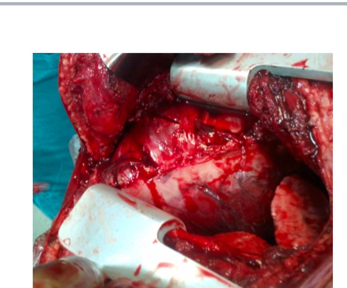
Left Poster lateral Thoracotomy Incision given, Layers deepened. Left pleura Opened in 6th ICS. All adhesions separated between left Lung and chest wall and Diaphragm. Left Lower lobe had two components, expanded lung and atelectic lung there was a systemic vessel supplying the left lower lobe from descending aorta near diaphragm. Systemic vessel triple ligation done and divided left inferior pulmonary vein and pulmonary arteries ligated and cut. Left inferior pulmonary Bronchus cut and sutured. Hemostasis achieved. Drains were kept. Ribs were closed with 5.0 Ethibond muscle Sutured with 2.0 vicryl. skin sutured with 3.0 Monocryl. Dressing done.
INTRAOPERATIVE FINDINGS
• Systemic artery started from the descending aorta and ending to left lower lobe.
• Partial expanded left lower lobe.
POSTOPERATIVE PERIOD
X-Ray PA view POD-1
X-Ray PA view POD-2
X-Ray PA view POD-3
Uneventful.
HISTOPATHOLOGY
Sections show collapsed lung parenchyma composed of few cystically dilated terminal and respiratory bronchioles lined by psuedostratified cilliated columnar epithelium, lumen filled micro abscess. Sub epithelium shows smooth muscle tissue, cartilage, lobules of mucus glands and mild infiltrates of neutrophils, lymphocytes and plasma cells. Focal lymphoid aggregates are seen the intervening alveoli are collapsed and lumen filled with denuded pnuemocytes, alveolar macrophages and hemorrhage. The interstitium shows moderate fibrosis thick walled vessels and dense infiltrates of lymphocytes, plasma cells, eosinophills, and macrophages. Focal lymphoid aggregates are seen. Also seen a medium sized thick walled blood vessel (systemic artery) which is adherent to the sequestrated lobe along with few(5) reactive lymph nodes.
DISCUSSION
Theories in the pathogenesis of pulmonary sequestration (5-15)
REFERENCES THEORY
1 1861 Rokitansky Fraction theory: separation of normally- developed lung.
2 1878 Ruge Excess theory: the malformation arises separately from the normal lung as the development of a rudimentary third lung.
3 1946 Pryce Traction theory: traction of an anomalous branch of the aorta on a developing lung segment separates it from the bronchopulmonary tree.
4 1956 Smith Blood supply theory: Insufficient arterial blood supply results in persistence of embryologic thoracic aortic arteries to the lung segment, with systemic blood pressure induced cystic degeneration of lung tissue.
5 1958 Boyden No causal relationship between nonfunctioning lung and systemic artery.
6 1959 Gebauer and Mason Theory of acquired anomalies: PS results from recurrent localized infectious process.
7 1967 Blesovsky Failure of embryologic growth factor signaling.
8 1968 Gene et al. Congenital bronchopulmonary foregut malformation theory: Accessory lung bud develops in the embryo and becomes either incorporated in the normal lung (Intralobar) or remains separated (extralobar).
9 1968 Morscarella and Wylie Intalobarsequestration is a collection of bronchogenic cysts associated with a systemic artery.
10 1984 Stocker and MalczakAn Acquired malformation with arterial supply derived from hypertrophy of pulmonary ligament arteries.
11 1987 Clements and warner Malinosculation theorya disruption the normal bronchopulmonary connection caused by an insult.
In the year 1946, Pryce coined the term sequestration. After that many theories of the pathogenesis of Pulmonary Sequestration have been proposed, while the most widely accepted hypothesis suggested that Pulmonary Sequestration was the consequence of an insult to the tip of developing lung buds. In early developing stages, the tips of the dividing bronchial buds are supplied by systemic capillary plexus derived from the primitive aorta. This plexus regresses with further lung maturation as the developing pulmonary artery takes over. The nature of the insult, probably more importantly the timing and severity of the insult, determines the morphology of the final lesions. Pulmonary Sequestrationis categorized into Intra Lobar Sequestration and Extra Lobar Sequestration. ILS is the most common form, constituting 92.5% of series from our hospital from 1990 to 2013 by Sun and Xiao With the development of diagnostic technology in China, increasing cases of PS, especially ELS, have been diagnosed correctly and given proper treatment in other local hospitals. Therefore, there were no ELS patients included in our series. It also suggests that ILS is more difficult to be distinguished. PS are manifested as a cough, purulent sputum, fever, etc. They all have symptoms similar to that of pneumonia. PS patients were susceptible to pulmonary infections. Without the presentation of pulmonary infection, PS is asymptomatic and only diagnosed incidentally as with 17.4% of patients in our study.
A number of case series on PS were reviewed along with this study. The characteristics of anomalous arteries described in this literature are summarized in . Few had done a pathological study for constituents of the vessel wall. The accompanying component of the anomalous systemic arteries in PS had not been studied in the previous publications. The systemic arteries of PS are potentially dangerous and should be identified before surgery. In this study, the aberrant systemic arteries were detected before surgery in 14 cases (70.0%) by contrast-enhanced CT (13/15, 86.7%) or CT angiography (4/4, 100%) and seen during surgery in the other six (30%), including four patients had taken CT noncontrast-enhanced before operation, and two patients (2/15, 13.3%) had taken contrast-enhanced CT. It is suggestive that a contrast-enhanced CT or CT angiography before the operation is essential for detecting potential abnormal arterial vessels. CT angiography is more useful to disclose PS. For patients with a pulmonary mass of unknown etiology, the possibility of PS should be considered preoperatively. A targeted observation of the systemic blood supply by enhanced CT or CTA is very important to reduce the risk of fatal intraoperative bleeding.
In PS, the aberrant arteries with elastic vessel walls represent a congenital anomaly, which cannot be reproduced by any form of interference to the pulmonary artery circulation. In dog models, in response to the ligation of the pulmonary artery, a large number of bronchial arteries with vessel walls of muscular nature were formed. Several investigators also indicated that the aberrant arteries in PS are distinct from bronchial arteries. A preponderance of elastic vessel walls in the aberrant arteries was identified in our study, consistent with the findings reported by Savic et al. This evidence could prove crucial in recognizing the congenital features of anomalous arterial supplies, as well as distinguishing it from bronchial arteries.
The aberrant systemic arteries of ILS are usually large in diameter. In Savic’s case series, the mean diameter values of the aberrant arteries were 6.3 mm (originating from the thoracic aorta) and 6.6 mm (originating from the abdominal aorta). The mean diameter values of the anomalous arteries in Yamanaka’s literature review were 9 mm at the origin, further dilating aneurysmatically (mean 20 mm in diameter) when entering the sequestrated lobe. In our study, the median diameter of the aberrant arteries was 8 mm, far exceeding the normal value of bronchial arteries. A relatively large diameter is also consistent with a congenital origin of the aberrant arterial supply. In our study, a majority of the aberrant systemic arteries originated from the descending aorta. Moreover, the origins of these aberrant arteries were close to the level of the sequestrated mass in the lung, leading them directly to the sequestrated region as a supply. This finding suggests that the aberrant artery and the sequestrated lung mass are closely related in position during embryonic development.
In general, pulmonary arteries and bronchial arteries go through the hilum in accompany with bronchi. Most of the aberrant systemic arteries examined in our cases penetrated directly the adjacent pleura to the sequestrated lung, without going through the hilum nor accompanied by any bronchial branch indicating that the aberrant arteries may originate from the systemic capillary plexus like the hypothesis, presenting a different way of evolution.
PROGNOSIS
Once intralobar sequestration is identified preoperatively andtreated properly by surgical ligation and resection of the lobe. Generally prognosis of the patient is good.
CONCLUSION
There are no certain pathology diagnostic criteria for the diagnosis of PS. The detecting of the aberrant systematic artery and distinguishing it from the bronchial arteries corresponded to certain lung abnormalities are the keys to the accurate diagnosis of pulmonary sequestration in adult patients. We propose that the characteristic features of the anomalous arteries include: Originating from aorta and its main branches, adjacent to the sequestrated area, directly running into the sequestrated mass without accompanying bronchus branch, being large in diameter, and having elastic vessel wall.
Key words: Aberrant systematic artery, congenital anomalous artery, pulmonary sequestration.


